
Imaging technology in radiation therapy uses medical imaging such as CT, MRI, or PET scans to create detailed pictures of the tumour and surrounding anatomy, which helps the treatment team accurately locate the target and plan, guide or verify radiation delivery.1-3
Why is imaging important?
Imaging plays an important role in the planning, simulation and delivery of radiation therapy.
Imaging technologies are used to locate and define the tumour and surrounding organs, and to support how radiation is delivered during treatment. It helps guide targeting of the treatment area and supports consistency between the planned and delivered treatment. In some cases, imaging information collected over the course of treatment may be used to adjust the treatment approach over time, this is called adapative radiotherapy.1–3
A range of imaging techniques may be used, including:1-3
- Computed Tomography (CT)
- Cone-beam CT (CBCT)
- Stereoscopic X-ray imaging
- Magnetic Resonance Imaging (MRI)
- Positron Emission Tomography (PET)
Advanced imaging may assist with:
- Identifying the tumour and surrounding organs
- Accounting for movement such as breathing or internal organ motion
- Monitoring anatomical changes over the course of treatment
- Verifying patient positioning before treatment sessions
1. Treatment planning (simulation)
Before treatment begins, an imaging scan (such as CT, MRI or PET) is performed to create detailed images of the tumour and nearby organs and tissues. These images are used by the treatment team to plan how radiation will be delivered.1,2,4–6
2. Imaging during treatment
Images may be taken before each treatment session to confirm the position of the tumour and surrounding anatomy. Some treatment systems, such as MR-Linac, allow detailed imaging to be performed during treatment delivery, providing visual information about tumour position and healthy surrounding tissues throughout the session.1,2,4–6
3. Monitoring and adaptation
Imaging collected during the course of radiation therapy allows the treatment team to monitor changes in anatomy and positioning. Where appropriate, this information may be used to adjust the treatment plan over time, called adaptive radiotherapy.1,2,4–6

Three main imaging types are typically used:
Stereoscopic X-Ray commonly used in CyberKnife treatment, is an early form of image guidance, developed in the 1990s. It offers a good level of accuracy for positioning and target verification; however, it may have limitations in soft tissue visualisation.3
In more recent years, more advanced 3D and real-time imaging technologies have become available, including:
- CBCT (Cone-Beam CT) – provides detailed 3D images of bones and surrounding soft tissues1
- MRI-guided imaging – tracks soft tissue in real time with high clarity1,5
CBCT technology is standard across all GenesisCare Linac machines and is used in IGRT (Image-Guided Radiation Therapy) and ART (Adaptive Radiation Therapy). Several GenesisCare locations also offer advanced MRI technology with high resolution soft tissue verification.
In stereoscopic X-ray imaging (the technology used for CyberKnife), primarily high-density objects are visible, such as bones and fiducial markers (for example, gold seed fiducial markers used in some prostate cancer treatments).3
In Image A (below), you can see the gold seed fiducial markers appearing white.
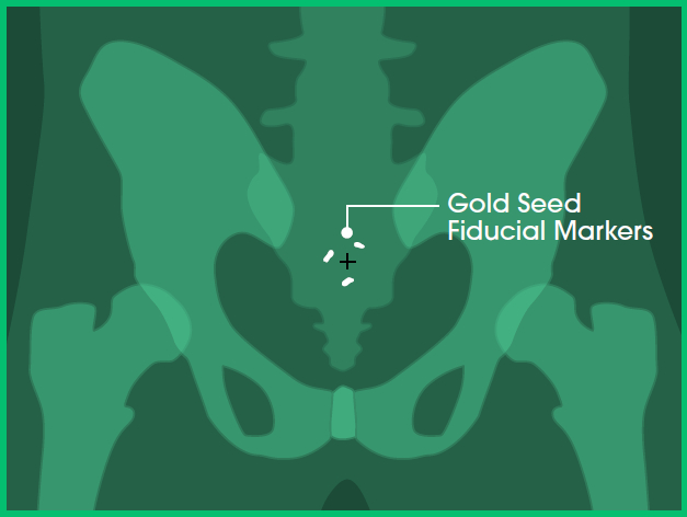
Image A: Front view
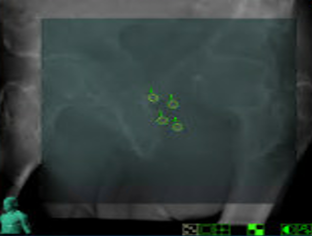
Image B: Clinical imaging scan
With cone-beam computed tomography (CBCT), it is possible to visualise a full three-dimensional alignment of the patient and tumour with surrounding anatomy.7
While high-density objects such as bones and fiducial markers remain visible (as in stereoscopic X-ray imaging), in addition CBCT also allows visualisation of soft tissues in three dimensions.8
In images A, B and C, you can see the:
- Prostate in red
- Bladder in blue
- Bowel in orange
- Fiducial markers in white
- Planned target volume outlined in yellow (where the radiation will hit)
In image D, which shows an example of CBCT imaging, you can see the:
- Planned target volume outlined in blue
- Bowel outlined in orange
This advanced level of imaging is standard of care used at GenesisCare and is available on linear accelerator (linac) machines for Image-Guided Radiation Therapy (IGRT) and Adaptive Radiation Therapy (ART).
Note: Certain Stereotactic Ablative Radiation Therapy (SABR) treatments cannot be performed with stereoscopic imaging alone. Examples include targeting tumours or nodes in the chest, abdomen, or pelvis where healthy structures need to be visualised before delivering treatment that stereoscopic imaging cannot see and avoid overdose of healthy tissues.9
Without sufficient visualisation of the tumour in relation to surrounding healthy organs, there is an increased risk of unintended radiation exposure to nearby tissue, which may be associated with side effects.8
When used in combination with fiducial markers, CBCT may also be used to monitor tumour position during treatment delivery by capturing images that visualise fiducials within the tumour. This allows the treatment team to assess positioning during treatment and respond if movement is detected.9
CBCT imaging is commonly used in the treatment of prostate, head and neck, lung and oral cancers.7
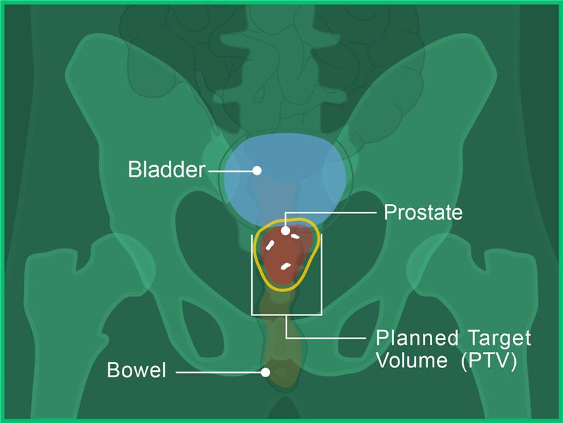
Image A: Front view
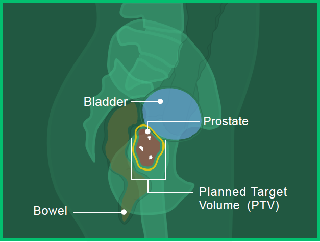
Image B: Side View
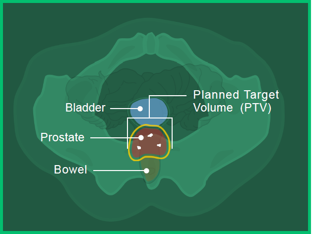
Image C: Bottom View
Image D: Clinical imaging scan
MRI enables high-resolution visualisation of soft tissue, in addition to high-density objects. It can also be used during treatment delivery to monitor movement.5
In image A, you can see the:
- Prostate in red
- Bladder in blue
- Bowel in orange
- Fiducial markers in white
- Planned target volume outlined in yellow (where the radiation will hit)
Image B shows the level of anatomical detail available with MRI-guided radiation therapy. You can see the:
- Planned target volume outlined in blue
- Bowel outlined in orange
MR-Linac may be used to treat various types of cancer, particularly those in areas that move with breathing or are located close to critical organs. Examples include liver, pancreas, adrenal, prostate, lung and rectal cancers.5
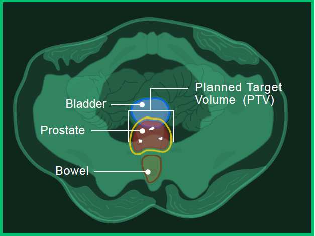
Image A: Bottom view
Image B: Clinical imaging scan
Imaging technology at GenesisCare
CBCT technology is standard across all GenesisCare Linac machines and is used in IGRT (Image-Guided Radiation Therapy) and ART (Adaptive Radiation Therapy). Several GenesisCare locations also offer advanced MRI technology with soft tissue mapping.
- Goyal, S., & Kataria, T. (2014). Image guidance in radiation therapy: Techniques and applications. Radiology Research and Practice, 2014, 705604. https://doi.org/10.1155/2014/705604
- Cancer Council Australia. (2023, March). Understanding radiation therapy. Retrieved September 2025, from https://www.cancer.org.au/assets/pdf/understanding-radiation-therapy-booklet
- Kurup, G. (2010). CyberKnife: A new paradigm in radiotherapy. Journal of Medical Physics, 35(2), 62–64. https://doi.org/10.4103/0971-6203.62128
- Radiation Oncology Targeting Cancer. (n.d.). Stereotactic ablative radiotherapy (SABR) in Australia and New Zealand. Retrieved September 2025, from https://www.targetingcancer.com.au/radiation-therapy/stereotactic-ablative-radiotherapy-sabr
- Hall, W. A., et al. (2022). Magnetic resonance linear accelerator technology and adaptive radiation therapy: An overview for clinicians. CA: A Cancer Journal for Clinicians, 72, 34–56. https://doi.org/10.3322/caac.21702
- Radiological Society of North America & American College of Radiology. (2024, June). Image-guided radiation therapy (IGRT). Retrieved June 2025, from https://www.radiologyinfo.org/en/info/igrt
- Mayo Clinic. (2023, November 30). Image-guided radiation therapy (IGRT). Retrieved September 2025, from https://www.mayoclinic.org/tests-procedures/image-guided-radiation-therapy/about/pac-20385267
- Radiological Society of North America & American College of Radiology. (2024, June). Image-guided radiation therapy (IGRT). Retrieved September 2025, from https://www.radiologyinfo.org/en/info/igrt
- Jaffray, D. A. (2012). Image-guided radiotherapy: from current concept to future perspectives. Nature reviews Clinical oncology, 9(12), 688-699.
Explore more

You are leaving our website
You are now leaving our website. GenesisCare do not control this content and therefore are not responsible for its accuracy or reliability.
You are leaving our website
You are now leaving our website. GenesisCare do not control this content and therefore are not responsible for its accuracy or reliability.